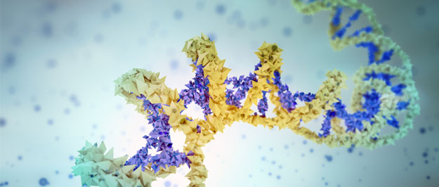
Circulating cell-free DNA (cfDNA) are small DNA fragments found circulating in plasma or serum, as well as other bodily fluids. The cfDNA isolated from plasma usually contains fragments of about ~170-500 bp, mostly corresponding to ~170 bp mononucleosomal and ~300 bp dinucleosomal DNA fragments [1,2], thought to arise mostly from apoptotic cells. In addition, larger fragments (>1,000 bp) are often detected, thought to arise mostly from necrotic cells. In healthy individuals, the levels of cfDNA in plasma/serum are generally low, ranging between ~0-50 ng/ml, however during pregnancy, illness, and exacerbation of tissue (intensive exercise or injury) the levels of cfDNA generally increase [3, 4].
The detection of increased levels of cfDNA during pregnancy and several diseases has gathered considerable attention for its potential application as a non-invasive method for diagnosis and monitoring of disease [3]. For example, during pregnancy cfDNA derived from the fetus is often present circulating in maternal plasma and has been utilized in pre-natal screening for trisomies and other fetal conditions [3]. In cancer patients, mutations in the Ras genes were among the first alterations reported in the cfDNA, and has been studied as a potential biomarker for monitoring tumor progression and recurrence, as well as response to therapy [5]. Other cfDNA alterations might have value for monitoring transplant rejection, diabetes, autoimmune, and cardiovascular diseases [3].
Epigenetic marks and clinical utility for diagnosis and/or monitoring of multiple conditions or diseases
Epigenetic alterations have also been detected in cfDNA [5], and two recent studies suggest that epigenetic marks could distinguish the tissue of origin of cfDNAs [6, 7]. Thus far distinction of the tissues contributing to the cfDNAs had relied on genetic differences between them, for example, comparing cfDNAs from tumor and normal, and comparing cfDNAs derived from fetus and mother. The detection of epigenetic alterations can be useful for diagnosing or monitoring multiple diseases that cannot be distinguished by genetic differences, or that don’t have distinct genetic features.
One of these epigenetic alterations is methylation. Lehmann-Werman and colleagues detected different methylation marks or patterns in cfDNA that could distinguish the tissue from which these cfDNAs originated [6]. First they identified tissue-specific methylation markers by examining publicly available data generated by Human Methylation 450 K arrays in tissue. Then they performed NGS of bisulfite-treated cfDNA from healthy individuals and patients of several pathological conditions, to assess the methylation of the identified markers specifically. For instance, the insulin gene (INS) promoter has previously been reported to be unmethylated in β-cells of type 1 diabetes patients, but methylated in other tissues. They were able to detect unmethylated INS promoter in cfDNA derived from β-cells of type 1 diabetes patients, whereas healthy individuals had undetectable or negligible amounts. They examined the cfDNA in patients suffering from other pathologies, and also found differential methylation that was detectable in the cfDNA of patients. Such changes include detection of unmethylated:
- INS promoter in β-cell derived cfDNA in type 1 diabetes patients
- MBP3 and WM1 loci in oligodendrocyte-derived cfDNA in multiple sclerosis patients
- Locus CG09787504 “Brain 1” in brain-derived cfDNA in patients with brain-damage
- REG1A and CUX2 genes in cfDNA derived from pancreatic cells in pancreatic cancer and pancreatitis patients
These findings have profound clinical implications, as they represent a promising non-invasive strategy for possible early detection and monitoring of many diseases, including pancreatic cancer, which up to date has no validated clinical biomarker for early detection, and is primarily detected at advanced stages, leading to a high morbidity and mortality rate.
Another epigenetic alteration is nucleosome positioning. Snyder et. al, reported that nucleosome maps, consisting of nucleosome and transcription factor occupancy, derived from the cfDNAs can distinguish the tissue of origin of the cfDNAs [7]. They performed deep sequencing of cfDNAs from healthy individuals and cancer patients, and found that the nucleosome maps derived from these were very distinct, and that the maps derived from the cfDNAs of healthy individuals correlated with hematopoietic cells, consistent with myeloid and lymphoid cells being the major contributing cells to the cfDNA in healthy individuals. Whereas the nucleosome maps derived from cfDNA of cancer patients contain cfDNA that correlated with non-hematopoietic cells, often correlating with cell types corresponding to same cancer type.
These findings could be useful for the classification of cancers during early diagnosis, and for a wider group of pathologies.
Challenges and limitations
Despite the extensive clinical utility of cfDNA, there are still some challenges in its study in the lab and its wide-spread clinical implementation. cfDNA is frequently found in plasma at very low concentrations, therefore its isolation and quantitation are not straightforward simple tasks. In addition, cfDNA with such alterations often compose just a small portion of the total cfDNA [8] circulating in plasma, and can be confounded by cfDNA contributed by normal cells.
Looking ahead
The above findings have profound clinical implications, as they represent a promising non-invasive strategy for the early detection and monitoring of numerous conditions. Based on detection of specific epigenetic alterations in the dying tissues contributing to the cfDNA circulating in plasma, it might be possible to diagnose disease even before the onset of clinical symptoms or progression to later, more advanced stages, where the disease burden becomes high, and is typically hard to manage or untreatable. Current studies could lead to the development and implementation of non-invasive blood-based diagnostic tests for a wide range of such diseases.
References
- Jahr, S., et al., DNA fragments in the blood plasma of cancer patients: quantitations and evidence for their origin from apoptotic and necrotic cells. Cancer Res, 2001. 61(4): p. 1659-65.
- Ivanov, M., et al., Non-random fragmentation patterns in circulating cell-free DNA reflect epigenetic regulation. BMC Genomics, 2015. 16 Suppl 13: p. S1.
- Swarup, V. and M.R. Rajeswari, Circulating (cell-free) nucleic acids–a promising, non-invasive tool for early detection of several human diseases. FEBS Lett, 2007. 581(5): p. 795-9.
- Devonshire, A.S., et al., Towards standardisation of cell-free DNA measurement in plasma: controls for extraction efficiency, fragment size bias and quantification. Anal Bioanal Chem, 2014. 406(26): p. 6499-512.
- Schwarzenbach, H., D.S. Hoon, and K. Pantel, Cell-free nucleic acids as biomarkers in cancer patients. Nat Rev Cancer, 2011. 11(6): p. 426-37.
- Lehmann-Werman, R., et al., Identification of tissue-specific cell death using methylation patterns of circulating DNA. Proc Natl Acad Sci U S A, 2016. 113(13): p. E1826-34.
- Snyder, M.W., et al., Cell-free DNA Comprises an In Vivo Nucleosome Footprint that Informs Its Tissues-Of-Origin. Cell, 2016. 164(1-2): p. 57-68.
- Danese, E., et al., Comparison of genetic and epigenetic alterations of primary tumors and matched plasma samples in patients with colorectal cancer. PLoS One, 2015. 10(5): p. e0126417.


