
Antibodies are used extensively in research for procedures, such as western blotting (WB), enzyme-linked immunosorbent assays (ELISA), and immunoprecipitation (IP). The issue of accuracy and reproducibility surrounding the usage of commercially available antibodies for research continues to be a concern within the scientific community.
The best way to compile information about an antibody is to review the provided manufacturer datasheet. In fact, some journals will not publish a study that includes an antibody with insufficient information and validation effort. Researchers must therefore carefully select the right antibodies for their experiments to avoid wasting valuable samples and funds.
Choosing a primary antibody
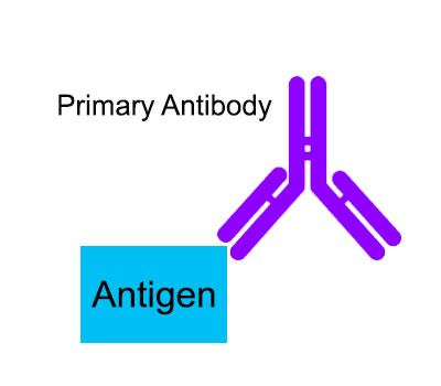
A primary antibody directly targets the antigen of interest. The species that the primary antibody was raised in should be different from the species of the sample. This mostly applies to samples with endogenous immunoglobulins (e.g., tissues that contain serum). Therefore, using an antibody that was raised against the same species as the experimental sample is most ideal so that non-specific binding is minimized.
Unless the datasheet specifies that the antibody has been validated for a species, there is no guarantee the antibody will work. However, if enough sequence homology is preserved, then an antibody may still be able to detect the same protein in different species. In this case, researchers will have to perform additional validation experiments.
Clonality of the primary antibody is an additional characteristic to consider. Although monoclonal and polyclonal antibodies have different advantages and disadvantages, both could potentially be suitable for an experiment.
An advantageous feature of monoclonal antibodies is that they possess a single epitope, so specificity is high and cross reactivity is minimized. Monoclonal antibodies are ideal for precision targeting. However, they are less likely to work across different species. Furthermore, epitope changes that occur in the target protein will also result in reduced binding. Monoclonal antibodies are produced from a single clone, which makes them relatively pure. However, the ascites fluid they are isolated from may still contain other antibodies that could affect specificity. Monoclonal antibodies are also typically more expensive because time consuming technical ex vivo processes are necessary to produce them.
Polyclonal antibodies are widely available, and at a lower cost. They are produced in vivo and are isolated from serum. They typically exhibit a range of detection. Because polyclonal antibodies possess multiple epitope protein recognition sites, they have a higher affinity to target proteins. This feature also helps with signal amplification and binding to target proteins even if epitope changes have occurred. However, the presence of multiple epitopes also increases cross-reactivity. Polyclonal antibodies are therefore less specific in comparison to monoclonal antibodies. To minimize this effect, companies have optimized their polyclonal purification processes. Blocking and other steps throughout the procedure can also be performed.
Choosing a secondary antibody

The secondary antibodies used in various assays are typically polyclonal. A secondary antibody should be raised against the primary antibody’s host being used. For instance, if a primary antibody is produced from rabbit, then the secondary antibody should be ‘anti-rabbit’. This information should be noted in the datasheet.
If you ‘re unsure about choosing your correct secondary antibody, please read How to Select the Right Secondary Antibody
After the antigen-primary antibody complex is formed, secondary antibodies are used for detection. The secondary antibody is often biotinylated, meaning it creates a strong bond with biotin-binding proteins, such as streptavidin. Streptavidin attached to reporter molecules can therefore be conjugated to secondary antibodies. Once a conjugated secondary antibody binds to the primary antibody, the signal is amplified. This is how antigen-antibody complexes can be visualized and quantified.
Fluorochrome antibody labels are typically used for flow cytometry and immunohistochemistry applications, which require a fluorescent microscope that can excite the dyes at different wavelengths. Chromogenic reactions occur when certain substrates are added to enzyme conjugated secondary antibodies. An example is Horseradish peroxidase (HRP), which is commonly used in colorimetric ELISAs. When TMB detection substrate is added to ELISA wells a blue colored precipitate forms. Western blotting with HRP is also performed, where immobilized antigen-antibody complexes appear as colored bands on the assay membrane.
Antibody validation for experiments
The datasheet should also specify what experimental conditions that antibody has been validated for. Although an antibody may have been produced with an affinity for a certain target, its binding specificity is still dependent on the procedure it is being used in. When an antibody that is not validated for a certain procedure is used, there is also a risk for false positive results. For instance, antibodies used for chromatin immunoprecipitation (ChIP) undergo stricter manufacturer requirements for purity and specificity. However, a ChIP-graded antibody may still work for a Western blot analysis.
Antibodies should be specific, selective and demonstrate reproducibility. The datasheet provides information about some validation processes, but it is not possible for a manufacturer to account for all different types of experimental variables. In addition to nonspecific binding and also the possibility of poor affinity between antibody and antigen, variability across different antibody lots can also be a cause for concern. Therefore, it is the responsibility of the investigator to validate all newly received antibodies.
Choosing controls are also an important step in the antibody validation process. A positive control cell, tissue or DNA sample should possess the target while a negative control sample, the target should be absent (knockout cells are also an option). Isotype-matched control immunoglobulin (IgG) is another commonly used control in immunoprecipitation. By treating a sample with an IgG antibody, an indication of assay background (i.e., nonspecific binding within the sample) can be determined. In order to test the reproducibility of an antibody, using multiple (or replicate) controls and repeating the experiment more than once is also recommended.
A western blot with various controls is commonly used for antibody validation. An antibody that is specific for a certain target protein will produce a band that has an expected protein molecular weight. Although this is a good first step, this only guarantees that the antibody can be used for a western blot analysis. If the actual experimental procedure is different, then additional validation tests within the context of the assay will likely be necessary.
References:
Couchman, JR. (2009). Commercial antibodies: The good, bad, and really ugly. J Histochem Cytochem, 57(1):7-8.
Saper, CB. (2005). An open letter to our readers on the use of antibodies. J Comp Neurol 493:477–478.
Bordeaux J., Welsh A., Agarwal S. et al. (2010). Antibody validation. Biotechniques. 48(3):197-209.